Micro Analysis and Visualization Lab (MAVL)
Lab Description
The Micro Analysis & Visualization Lab (MAVL) is an Institute-wide facility for microscopic analysis and imaging services. The MAVL provides researchers with access to a range of microscopic platforms including low-magnification stereozoom reflected-light (dissecting) scopes, transmitted-light compound scopes in a variety of configurations (including petrographic and fluorescence capabilities), inverted scopes for live cell or tissue analysis, and a high-magnification tabletop scanning electron microscope equipped for elemental composition analysis. Microscope cameras and basic image analysis software are available for many of the platforms. The MAVL was created in 2015 as a dedicated research space, equipment library and teaching/training facility that all at DRI can use, contribute to and share.
Equipment from the MAVL can be used by DRI researchers for projects such as characterization of pollutants in air and water samples, particle morphology and elemental analysis, identification of microfossils, or forensics. To discuss research needs related to micro-imaging and analysis, contact lab manager Dave Rhode at dave.rhode@dri.edu.
Photo Gallery
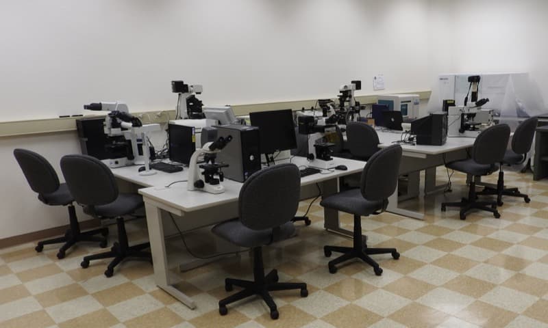
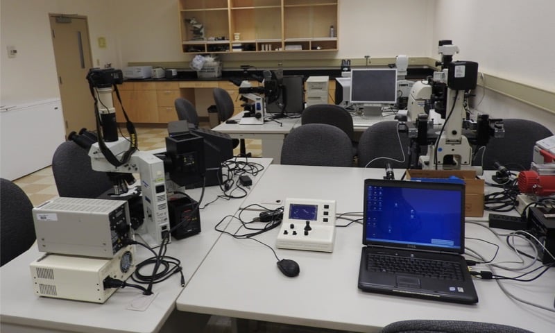
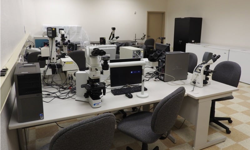
MAVL-workstation
Work stations in the MAVL can be configured to your specialized needs.
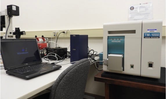
MAVL-hitachitm-1000
Hitachi TM-1000 tabletop scanning electron microscope, capable of 25-10,000X magnification. This unit is equipped with a Swift energy dispersive spectroscopic unit for elemental micro-characterization.

MAVL-willow-charcoal
An image obtained from the Hitachi TM-1000: Willow charcoal.
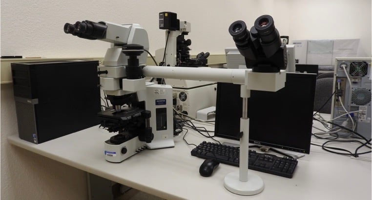
MAVL-olympus-bx51
Olympus BX-51 transmitted compound microscope, with side viewing attachment for teaching
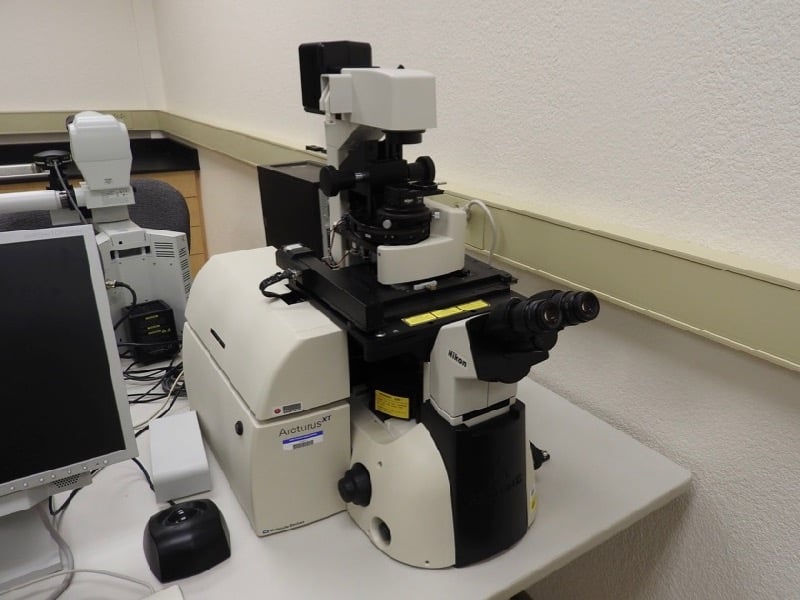
MAVL-nikon-eclipse-arcturis-laser
Nikon Eclipse inverted scope equipped with Arcturus laser microdissection system for cell capture and analysis.
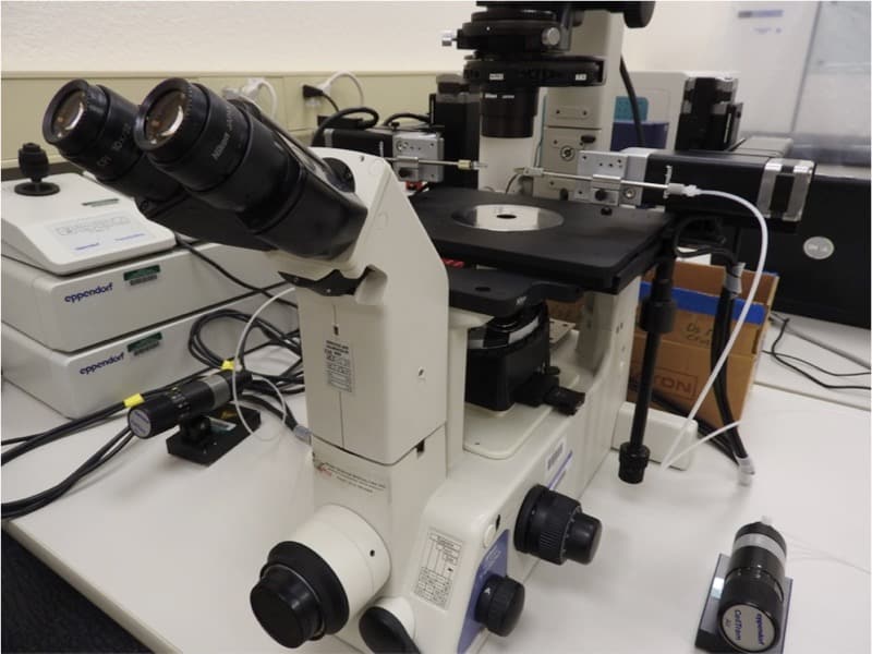
MAVL-nikon-eclipse-eppendorf-micromanipulators
Nikon Eclipse inverted scope equipped with Eppendorf micromanipulators for live cell/tissue sample examination.
MAVL-maxey-science-building
The MAVL is located in DRI’s Maxey Science Building, Room 148.
MAVL-equipment
To use the MAVL or to learn more about the equipment, please contact Dave Rhode.
CONTACT
Dave Rhode, Ph.D.
Lab Director
Dave.Rhode@dri.edu
LAB LOCATION
Desert Research Institute
2215 Raggio Parkway
Reno, NV 89512
DIVISION
Earth & Ecosystem Sciences